Pseudocyst
In this article, we will discuss briefly the types of Pseudocysts that effect the jaws, the clinical pictures, radiographic appearance and treatment. If intrested in the other types of cysts of the Jaw, please click the the following links:
[divider scroll_text=””]
Aneurysmal Bone cyst
Features
- It is called Psuedocysts because they appear Radiographically as cyst-like lesion but microscopically exhibit no epithelial lining.
- It may occur alone or associated with other disorders of bone.
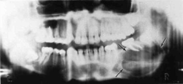
Aneurysmal bone cyst producing expansion of the cortical plates - Benign lesion; in maxilla , mandible or any other bones.
- It is a rare non-neoplastic lesion of bone of unknown etiology.
Clinical Features
- Younger than 30.
- It affect maxilla and mandible especially in their posterior region
- It produces firm, diffuse swelling with facial asymmetry due to expansion of the bone.
- May be Associated with Pain.
- The cyst expands slowly, and sudden expansion following minor trauma is indicative of pain and bleeding inside the cyst.
Radiographically
- It may appear as a unilocular radiolucency, some cases appear as a multilocular radiolucency.
- The teeth may displaced, show some root resorption but are still vital
Treatment
- A relatively High recurrent rate with simple curettage.
- Excision or curettage with supplemental cryotherapy is the treatment of choice.
[divider scroll_text=””]
Simple Bone cyst
Traumatic Bone cyst, Hemorrhagic bone cyst, Unicameral bone cyst

Features
- Empty Intrabony cavity that lacks an epithelial lining.
- The pathogenesis is not Known, It has been suggested that trauma to the bone produces intramedullary haemorrhage which fails to organise and heal and that cavitation occurs by subsequent hemolysis and resorption of the clot.
- Teenagers are most commonly affected.
- Mandible is the most common side.
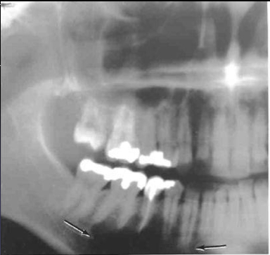
Clinical Picture
- Simple Bone Cysts are very common lesions.
- Most occur in the first two decades of life, with a mean age of 17 years.
- The lesion shows a male predominance of approximately 2:1.
- The cyst arises most frequently in the premolar and molar regions of the lower jaw.
- The majority of solitary bone cysts are symptomless with some degree of bony expansion in some cases.
Radiographically
- Internal structures are totally radiolucent.
- Commonly in the Mandible especially in the Ramus of the mandible.
- Well demarcated unilocular or multilocular radiolucency with scalloping around roots commonly found in posterior mandible.
- Displacement of teeth is uncommon.
Treatment
- Surgical exploration of the area will confirm the diagnosis. Opening such cysts, evacuation of their contents and making them bleed results in healing of the cavity.
- Some cases may heal spontaneously
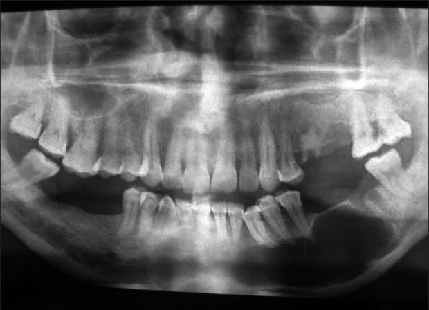
Simple Bone cyst
Differential Diagnosis
Simple bone Cysts may have an appearance similar to that of a true cyst, especially an Odontogenic Keratocyst (Primordial Cyst). However, maintenance of some lamina dura and the lack of an invasive periphery and bone destruction should be enough to remove this category of diseases from consideration.
[divider scroll_text=””]
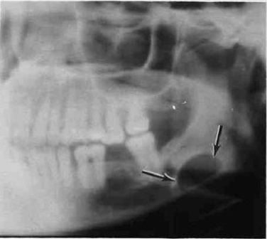
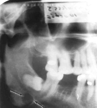
Static bone cyst
Stefan’s bone defect, Developmental salivary gland defect, latent bone cyst
Feature
- A developmental salivary gland defect of the mandible
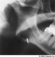
Static bone cyst - The defect contains either salivary gland or adipose tissue.
- The cavity occurs due to a developmental (deep and well-defined) depression defect on the lingual aspect of mandible which contains a portion of sublingual or submandibular salivary gland.
Clinical Picture
- Asymptotic. It is found by coincidence during routine radiographic examination which occurs between the premolars and the angle of the jaw.
- It is Located below Mandibular canal in molar area. More Accurately, The most common location is within the submandibular gland fossa and often close to the inferior border of the mandible.
Radiographically
- This is an uncommon developmental anomaly of the lower jaw which may be mistaken as a cyst on x-ray.
- sharply circumscribed oval Radiolucency beneath Inferior Alveolar Canal, with encroachment on inferior border of the mandible.
- Shape is round, ovoid, or, occasionally, lobulated radiolucency that ranges in diameter from 1 to 3cm Below the inferior alveolar nerve canal anterior to the angle of mandible, in the region of the antegonial notch.
- The margins of the radiolucent defect are well defined by a dense radiopaque line, Discrete corticated margin.
- This cortical margin usually is thicker on the superior aspect.
Treatment
- No biopsy or Treatment is required.
[column col=”1/2″]
[/column][column col=”1/2″ last=”true”]
[/column][divider scroll_text=””]
Sources
- Ibn Sina University Curriculum By Dr. M. Shanbari.
- Intelligent Dental
OziDent Members Only
The rest of article is viewable only to site members,Please Register and/ or Confirm registration via EmailHere.If you are an existing user, please login.