In this article, we give in details the facts, factors, pathogenesis, clinical picture, diagnosis, treatment of Pemphigus Vulgaris. A type of Autoimmune disease effecting the skin and oral mucousa.
Pemphigus Vulgaris
Facts
- Its autoimmune disease involving skin, mucous membrane showing vesicles and bullae.
- Uncommon and fatal.
[divider scroll_text=”SCROLL_TEXT”]
Etiology and Pathogenesis
- Characterised by circulating IgG autoantibodies against intercellular cementing substances in the epithelium. (Desmoglein)
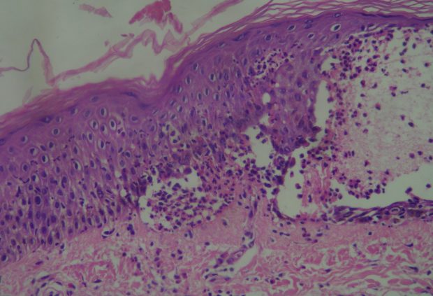
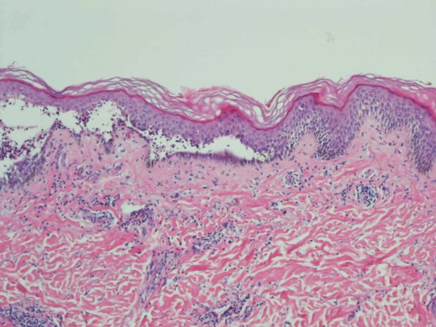
Suprabasial acantholysis - Once the autoantibodies attaches to the Antigen (cementing substance)
- Release of proteolytic enzmes from epithelial cells
- Destruction of the intercellular cementseparation of the epithelial cell from each other (acantholysis).
- Presence of the weakest junction supra‐basal
- Split between the basal and prickle layer (supra‐basal cleft)
- Accumulation of fluid in the supra‐basal clefts
- Formation of epithelial vesicle and bullae.
[divider scroll_text=”SCROLL_TEXT”]
Clinical Picture
- Age: 40 – 60 years old.
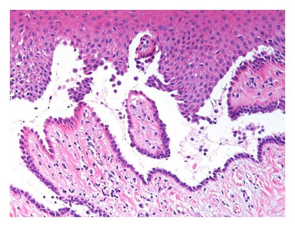
Suprabasial acantholysis near the tips of two adjacent rete pegs is recognized. - Sex: Female.
- Site: Skin and Mucous Membrane.
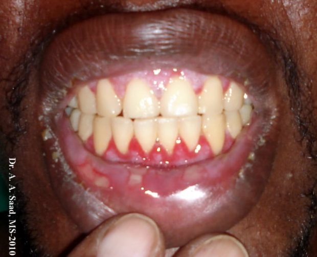
Oral Features
- Site
- Positive Nikolsky Sign
- The Bullae/ vesicle
- Thin walled on a non‐ erythematous base.
- The bulla rapidly ruptures and breaks giving a shallow ulcer.
- The ulcer is irregular with detached margins on the peripheral, why? The edges of the ulcer continue to extend peripherally thus increasing in size (extend to lip crust forming) + (extend to the throat leads to difficulty in swallowing) leading to detached epithelium at the margins and irregular margins.
- The ulcers are: Big, irregular, shallow and bleeding, why? Because the ulcers has epithelium detachments on the peripheral.
[divider scroll_text=”SCROLL_TEXT”]
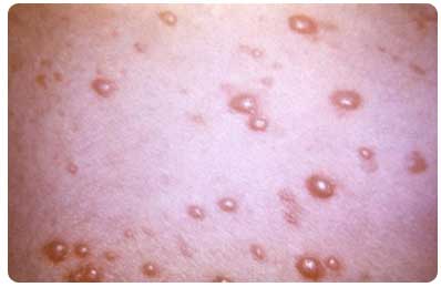
Skin Features
- Site: Groin, Axilla, face and neck.
- The Bullae/ Vesicle: Thin walled on a non erythematous Base, Shallow ulcers and contains clear or Hemorrhagic or seropuruelnt fluid.
- The bullae ruptures leading to ( ulcers + bleeding + easy detachment of epithelium on the periphery) thus the ulcers have an irregular margin. Why irregular margins of the ulcers? Because of easy detachment of epithelium on the periphery of the ulcer.
- Postive Nikolksy Sign.
[divider scroll_text=”SCROLL_TEXT”]
Nikolsky Sign
Nikolsky’s sign is a clinical dermatological indicator, named after Pyotr Nikolsky. The sign is shows when slight rubbing of the epidermis results in exfoliation of the outermost layer forming a blister within instantly. This sign is almost always present with toxic epidermal necrolysis and is associated with pemphigus vulgaris.
Nikolsky’s sign is positive in Pemphigus Vulgaris and shows in the following Points:
- On intact oral mucosa: Lateral pressure may lead to peeling of epithelium leaving a large denuded area (Desquamated Gingivitis) or formation of vesicle or bullae.
- On vesicle: Vertical pressure extension of lesion to adjacent tissue increase in size of vesicle.
- On Skin:
- Pressure on the Outer layer of epithelium is easily removed and slipped.
- Pressure on skin will lead to formation of bullae or vesicle.
- Pressure on an intact vesicle will lead to forcing the fluid to the surrounding unaffected tissue.
[divider scroll_text=”SCROLL_TEXT”]
Diagnosis
- Case History.
- Clinical Examination by a Postive Nikolsky’s Sign.
- Laboratory Investigations.
Laboratory Investigations
- Biopsy : Under microscope, Intra-epithelial Bullae is present.
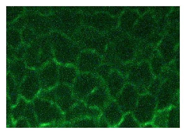
Immunofluorescence for deposits of IgG - Immunofluorescence: to help detect the presence of IgG in the inter-cellular attachment zone.
- Direct Results: detects the tissue with the IgG autoantibody attached to them.
- Indirect Results: detects the presence of Autoantibody circulating in the blood.
- Cytologic Smear Showing T-Zank Cells.
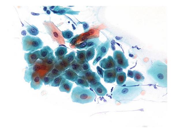
Acantholytic Tzank cells.
[divider scroll_text=”SCROLL_TEXT”]
Treatment
- Treatment is based on Steroids , pain management and good hygiene.
- Early diagnosis improves outcome of treatment, why? At later stage severe skin involvement will lead to secondary infection and severe imbalance of fluid electrolyte, this is considered fatal. In this state high dosage of corticosteroid is used to control the disease. The amount of corticosteroids used should be monitored, why? because risk of death due to high dosage of corticosteroids is riskier than the disease it self.
- Topical Drugs for the oral lesions include Topical Anaesthesia, Antiseptics and Corticosteroids.
- Systemic corticosteroids: Used alone or combined with immune‐suppressive drugs, why combined? To reduce the dosage, e.g. Azathioprine, Cyclosphosphamide.
[divider scroll_text=”SCROLL_TEXT”]
Sources
- Hindawi.com Journal.
- Misr International University By Dr. Mohsen S. Mohamed.
- Med India.
- Derma Rounds
- Wikipedia.
OziDent Members Only
The rest of article is viewable only to site members,Please Register and/ or Confirm registration via EmailHere.If you are an existing user, please login.
