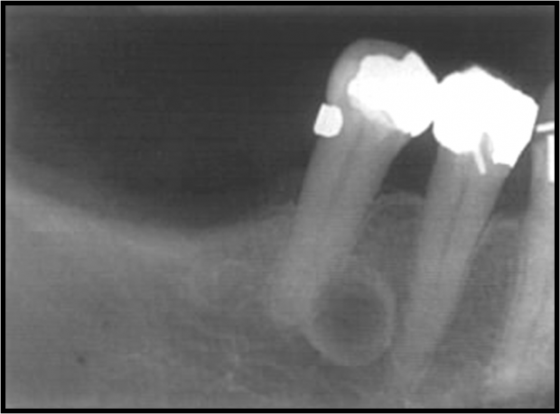
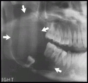
Odontogenic Cysts
Radicular cyst
Other Names
Periapical cyst, apical periodontal cyst, or dental cyst.
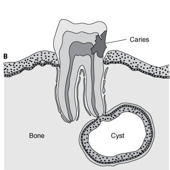
Features
- Most common.
- Inflammatory cyst because in the majority of cases it is a consequence to pulpal necrosis with an associated periapical inflammatory response. ( Rests of Malassez )
- Related to the apex of non vital tooth or related to lateral canal.
- cyst formation occurs as a result of epithelial proliferation, which helps to separate the inflammatory stimulus ( necrotic pulp ) from surrounding bone.
Clinical features
- Asymptomatic, unless associated with infection or intra-cystic pressure.
- Swelling of the jaw is the chief complaint.
- Soft fluctuant swelling, bluish in color beneath the mucosa.
- Cause bone resorption.
- On palpation may feel bony and hard unless if bone resorption occurs thus it feels soft and rubbery.
- Slight Male Prevalence.
- Occurring in 3rd to 6th decade.
- 60% in Maxilla Canine or incisors.
Radiographically
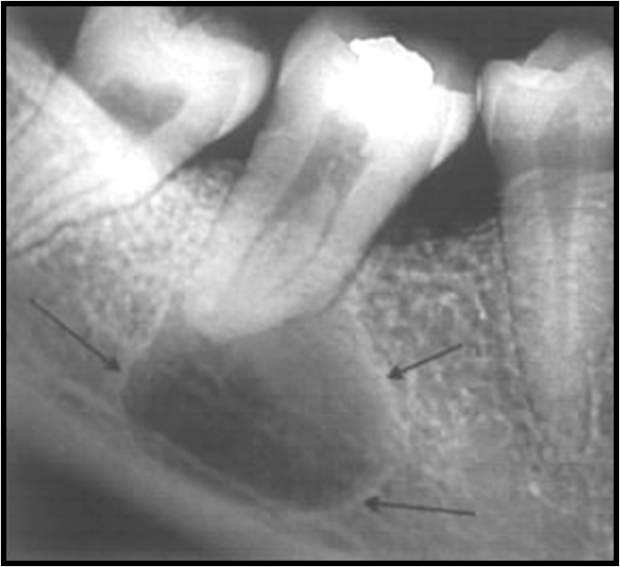
- Location apex of a non-vital tooth, but occasionally on the mesial or distal of the teeth.
- Cannot be differentiated from periapical granuloma.
- Radiolucency ( round or ovoid ) with narrow, opaque margin that is continuous with the lamina Dura.
- well defined cortical border.
- Root Resorption of offending tooth and occasionally the adjacent tooth.
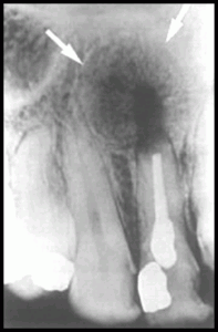
Differential Diagnosis
- Odontogenic keratocyst.
- Stafne’s developmental salivary gland defect.
Treatment
- Extraction of the associated non-vital tooth and curettage of the apical zone.
- RCT may be performed with an apicoectomy ( direct curettage of the lesion ).
- The most often used is only RCT, since most periapical lesions are granulomas and resolve after removal of the inflammatory stimulus ( necrotic pulp ).
- Surgery ( apicoectomy and curettage ) is done for persist lesions, presence of a cyst or inadequate RCT. Residual cyst may occur if the cyst lining incompletely removed.
Residual cyst
Features
- It arises either due to improper surgical elimination of a radicular cyst or extraction of the related tooth without removal of the cyst
Clinical Features
- Asymptomatic and often is discovered on radiographic examination of an edentulous area.
- Its clinical and histological characteristics are identical to those of a radicular cyst
- Radiographically it will seen as a radiolucency of variable size at the site of a previous tooth extraction.
Radiographically
- Often in the mandible and always above the inferior alveolar nerve canal.
- Periphery and shape. cortical margin shape is oval or circular.
Differential Diagnosis
- Odontogenic keratocyst.
- Stafne’s developmental salivary gland defect.
Treatment
- complete removal of the cystic lesion ( enucleation ).
- In large cyst; Marsupialization is achieved by creating a window to evacuate the cyst and to decrease the intracystic pressure. Refer to Article to explain the Difference between.
Lateral periodontal cyst
Features
Clinical Features
- Asymptomatic.
- Mandibular premolars and bicuspid region and occasionally in the incisors areas.
- Maxillary lateral incisors region.
- 4th and 5th decades of life most effected.
Radiographically
- Well-delineated, round or teardrop-shaped.
- Unilocular radiolucency with opaque margin along the lateral surface of a vital tooth root.
Diffrentail Diagnosis
- A small odontogenic Keratocyst.
- Small mental foramen.
- small neurofibroma
- a radicular cyst at the foramen of a lateral (accessory) pulp canal.
Treatment
- Local excision is generally curative.
- In case of multilocular ( botryoid ) cyst seems to have recurrence potential, follow up, therefore suggested for treated multilocular odontogenic cyst.
Gingival cyst ( Newly Born)
Features
- It is a cyst that arises from the rest cells of the dental lamina
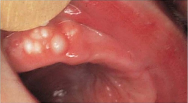
- Solitary nodules on the crest of the alveolar ridge of the new born or very young infants. it appears as a multiple, small, firm, white or grayish-white nodules on the alveolar ridge .
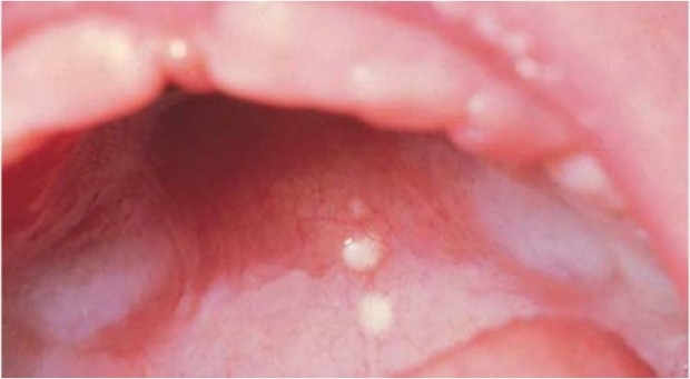
- Epstein´s pearls are cystic nodules found on the hard palate or at the junction of hard and soft palate.
Treatment
- No treatment is necessary, because spontaneous rupture usually occur early.
Gingival cyst ( Adults)
Features
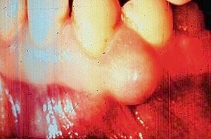
- A small developmental cyst of the gingival tissues derived from the remnants of dental lamina. It appears as a small circumscribed painless swelling, same color as the color of the gingiva.
Treatment
- Local excision.
Epstein’s pearls
Bohn’s nodules
Dental lamina
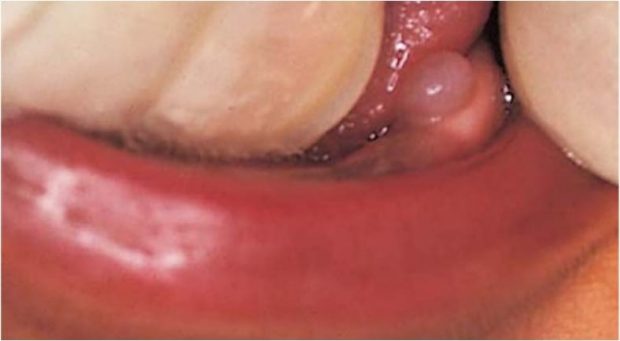
Dentigerous or follicular cyst
Definitions
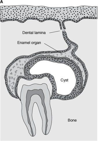
Dentigerous Cyst is one which encloses part or all of an unerupted crown of a tooth. It is attached to the junction where the tooth enamel meets the root (amelocemental junction) and arises in the follicular tissues covering the fully formed crown of the unerupted tooth.
Features
- Second most common.
- Arise from reduced enamel epithelium of the dental follicle of an unerupted tooth.
- Developmental.
- Attached to the tooth cervix ( ECJ ).
- Enclose the crown of unerupted tooth.
Clinical Features
- Associated with 3rd molar and maxillary canine; mostly IMPACTED.
- More in Mandible.
- 2nd to 3rd decades.
- More in Males
Radiographically
- Well-defined, unilocular or occasionally multilocular Radiolucency.
- The unerupted tooth is often displaced.
- n the mandible the RL may extend to the ramus or to the body of mandible.
- In maxillary canine region the cyst may extend into the sinus or to the orbital floor.
- Resorption of adjacent tooth root maybe seen.
Treatment
- Extraction of the tooth and enucleation.
- Marsupilization of the cyst to allow for decompression and subsequent shrinkage of the lesion.
Eruption cyst
Definition
The eruption cyst is the soft tissue analogue of the follicular cyst.
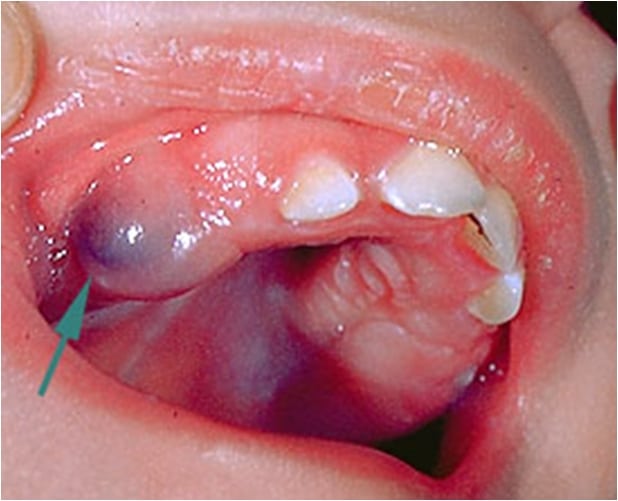
Features
- Developmental.
- Result from fluid accumulation within the follicular space of an erupting tooth.
- The epithelium lining this space is simply reduced.
Clinical Features
- It appears as soft fluctuant blue to dark red mass on the alveolar ridge in place of any erupting tooth, particularly molar and canines.
Radiographically
- Negative as it is soft tissue cyst above the crown of unerupted tooth.
Treatment
- No treatment is needed, because the tooth erupts through the lesion, the cyst disappears spontaneously without complication.
Odontogenic Keratocyst (Primordial Cyst)
Features
- Arises from reduced enamel epithelium, dental lamina rests and malassez rests.
- May exhibit aggressive clinical behavior.
- Associated with nevoid basal cell carcinoma.
- It can be found any where in the jaw.
- Highly recurrence after initial surgical intervention.
- characterized by keratinizatoin and budding cyst lining.
- Has a wide age range but there is a peak incidence in the 2nd and 3rd decade with a smaller peak in the 5th decade.
- The cysts are more common in males and 70% occur in the wisdom tooth and ramus region of the lower jaw.
- Can Reach Large sizes without causing expansion, because it predominantly enlarges anterior-posterior direction.
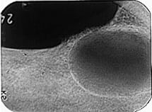
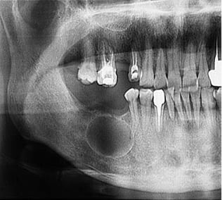
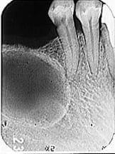
Clinical Features
- Arises from reduced enamel epithelium, dental lamina rests and malassez rests.
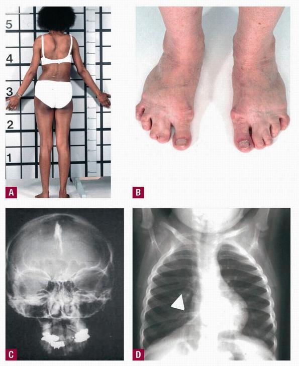
Gorlin syndrome Manifestations - May exhibit aggressive clinical behavior.
- Associated with nevoid basal cell carcinoma. (Gorlin syndrome) it has many manifestations:
- Oral – multiple odontogenic keratocysts, cleft lip or palate.
- Skin – multiple nevoid basal cell carcinoma
- Skeletal – rib anomalies, vertebral deformities, polydactyly (Birth defect characterized by the presence of more than the normal number of fingers or toes)
- Central nervous system – calcified falx cerebri, brain tumors
- It can be found any where in the jaw.
- Highly recurrence after initial surgical intervention.
- Characterized by keratinizatoin and budding cyst lining.
Radiographically
- Well-circumscribed Radiolucency with smooth radiopaque margins.
- Multilocular most common in large lesions.
- It may cause buccal expansion.
- It may cause displacement of roots or IAC.
- Resorption of the root is common.
- May be associated with missing or unerupted tooth.
- It maybe difficult to distinguish from Dentigerous cyst.
Differential Diagnosis
- Dentigerous cyst
- The cyst is likely to be an OKC if the cyst is connected to the tooth at a point apical to the cementoenameljunction or if no expansion of the cortical plates has occurred.
- Ameloblastoma.
- Odontogenic myxoma.
- A simple bone cyst.
Treatment
- Surgical excision with peripheral osseous curettage or osteoectomy is the preferred method of management.
- The use of chemical cauterization of the cyst in Highly recurrence cyst has been advocated.
- In Large cyst; Marsupilization, followed by enucleation, may be an attractive alternative.
Reasons for recurrences
- Thin and fragile cystic epithelium.
- Has satellite cysts (daughter cysts).
- Epithelial lining has intrinsic growth potential.
- Has increased mitotic activity.
- Patients with nevoid basal cell syndrome have particularly tendency for recurrence of keratocysts.
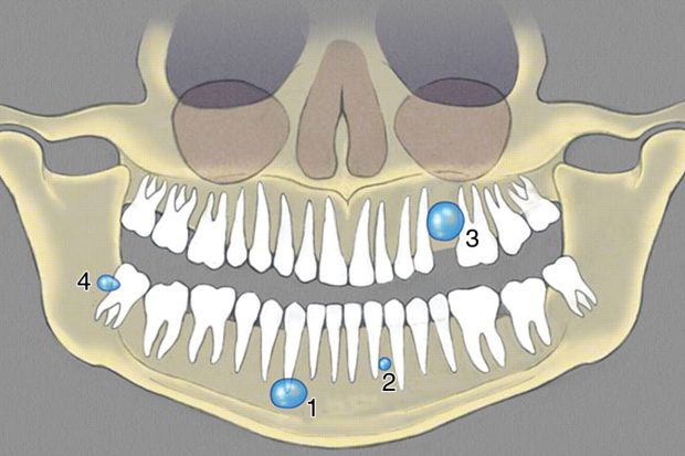
Glandular Odontogentic Cyst
Features
- Rare.
- Developmental odontogenic cyst.
- Most common site of occurrence is the front region of the lower jaw.
- They present as slow-growing, painless swellings.
- The cyst has a potentially aggressive, locally invasive nature.
- Recurrence tendency.
Paradental Cyst
Features
- Arises along partly erupted wisdom teeth that is involved by pericoronitis.
- Mostly in the lower jaw.
- Located at the cheeks side of the tooth
Sources
- Ibn Sina University Curriculum By Dr. M. Shanbari.
- INTELLIGENT DENTAL RADIOGRAPHIC APPEARANCE OF CYSTS PART 2 by Dr. MEIFONG.
- Wikipedia.
OziDent Members Only
The rest of article is viewable only to site members,Please Register and/ or Confirm registration via EmailHere.If you are an existing user, please login.
